Imaging of the Hand & Wrist

Hand & Wrist

Imaging Recommendations
Osteoarthritis in the hand and wrist is associated with increasing age. It typically occurs in the basal joint of the thumb or distal interphalangeal joints and is more commonly diagnosed in women. In the majority of cases clinical examination is as good as imaging for diagnosis.
X-rays show it is present in about 25% of women over the age of 55 but some of these may be asymptomatic.
Xrays are recommended for Trauma and can sometimes be useful in suspected erosive arthritis (eg gout or rheumatoid) or Osteoarthritis
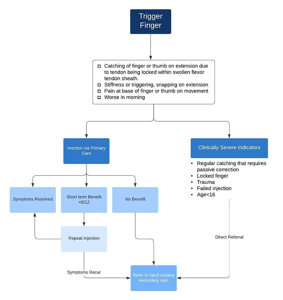
Ultrasound can be useful to support a clinical diagnosis of De Quervain's tenosynovitis or to guide injections of steroid such as in carpal tunnel syndrome or trigger finger. Aspiration of ganglions can be done in primary care and US-guided ganglion aspirations are not funded by the CCG. Any referral made for this will be returned unless exceptional circumstances.
There are no routine indications for wrist or hand MRI imaging from primary care. Any referral for these should be discussed with a musculoskeletal radiologist for approval.
Patient information for the following conditions and others can be found at the British Society Hand Surgery Website